Development of Microfluidic Chio Capable of Verifying Drug Resistance of Cancer Cells Through In Vitro Culture
Professors Seok Chung and Jason K. Sa’s group at KU conducted the joint study with Professor Hye Won Lee of the National Cancer Center.
The research results were published in Advanced Science, a globally renowned journal.

▲(From left) Dr. Hyunho Kim (first author), Professor Jason K. Sa (first author), Professor Hye Won Lee (corresponding author) and Professor Seok Chung (corresponding author)
Professor Seok Chung’s group of the Department of Mechanical Engineering in the College of Engineering conducted a joint study with Dr. Hyunho Kim of the Harvard Medical School, Professor Jason K. Sa of the College of Medicine at KU and Professor Hye Won Lee of the National Cancer Center. They developed a microfluidic chip capable of verifying the resistance of cancer cells to anti-cancer drugs by culturing a patient’s cancer cells with their adjacent cells. Dr. Hyunho Kim is now taking a postdoctoral course at the Harvard Medical School, and Professor Hakho Lee of the Harvard Medical School also participated in the present study. The research results were published online in Advanced Science (IF: 16.8), a globally renowned journal, on June 3.
* Article Title: Recapitulated crosstalk between cerebral metastatic lung cancer cells and brain perivascular tumor microenvironment in a microfluidic co-culture chip
Providing patient-customized treatment requires the selection of drugs appropriate to the genomic information of the patient’s cancer cells, the characteristics of those cancer cells, and the environment of the cancer tissue. However, due to the genetic diversity of tumors, genomic information alone can barely provide suitable targeted therapies. In addition, animal tests performed using mice often fail to represent diverse human cells and tumor microenvironments. Overcoming these limitations necessitates using human cells to reconstruct the features of the original tumor environment in which the cancer cells grow.
In the present study, microfluidic chip technology was employed to allow the cancer cells separated from a cancer patient to grow in their specific microenvironment. The microfluidic chip developed in this study was particularly targeted to metastatic lung cancer cells located in the brain because the uniqueness of the brain often prevents the conventionally administered drugs from functioning. Unique features of the brain microenvironment, such as the blood brain barrier or astrocytes, form a mechanism that protects cancer cells.
The research team reconstructed the brain microenvironment by culturing a microenvironment consisting of cerebrovascular cells, astrocytes and extracellular matrix in a microfluidic chip. In addition, the team successfully performed a coculture with cancer cells derived from a patient with brain metastatic lung cancer. The drug response of a cancer cell culture only and the drug response of a co-culture with the microenvironment was compared using genome sequencing and molecular profiling. The comparison revealed the changes in the lung cancer cells caused by the brain microenvironment.
Compared with the existing cell culture platform, the microfluidic chip has a short intercellular interval of micrometers allowing the concentrations of cytokines (or chemical factors) secreted from cells to rapidly increase and be maintained in the channel, thereby realizing close-knit intercellular signal transmission. In this way, the microfluidic chip can recapitulate a process in which cancer cells and microenvironmental cells are assimilated with each other as in vivo.
The anticancer drugs used in clinical settings were applied to the chips, and the results showed that the drug responses differed from the genomic predictions. When the lung cancer cells were cultured in the brain microenvironment, the cancer cell survival rate increased and the transcriptional network profile changed. This process may approximate the drug resistance factors formed in the bodies of actual patients.
The research team commented, “We developed a nonclinical study platform that can reconstruct the interaction between cancer cells and their surrounding microenvironment. This interaction plays the principal role in contributing to the resistance against existing cancer therapies. This platform will be used to investigate the molecular biological mechanisms of the cancer cell-paracancerous microenvironment ecosystem and prepare new anticancer treatment strategies.”
The study was supported by the Mid-Career Research Program and the Bio & Medical Technology Development Program of the National Research Foundation (NRF), the 3D Biochip Based Drug Development Platform Program of the Ministry of Trade, Industry and Energy, and the Korea Health Technology R&D Project of the Korea Health Industry Development Institute.
* Article Title : Recapitulated crosstalk between cerebral metastatic lung cancer cells and brain perivascular tumor microenvironment in a microfluidic co-culture chip
* Journal : Advanced Science
* Authors: Dr. Hyunho Kim (first author/KU), Professor Jason K. Sa (first author/KU), Jaehoon Kim (coauthor/KU), Dong-Hee Choi (coauthor/KU), Seok-Hyeon Kang (coauthor/KU), Hyun Jeong Oh (coauthor/KU), Da Eun Jeong (coauthor/Thermo Scientific), Do-Hyun Nam (coauthor/Samsung Medical Center), Hakho Lee (coauthor/Harvard Medical School), Hye Won Lee (corresponding author/National Cancer Center) and Seok Chung (corresponding author/KU).
Professor Hye Won Lee (corresponding author) and Professor Seok Chung (corresponding author)
[ Figures ]
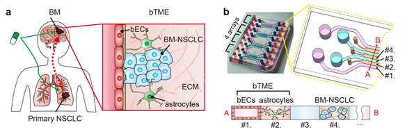
▲ Figure 1 a) Illustration of the environment of the cancer cells from a patient with brain metastatic lung cancer. The environment primarily consists of cancer cells, cerebrovascular cells, astrocytes and extracellular matrix; and b) The microfluidic chip developed in the present study to recapitulate the microenvironment of brain metastatic cancer cells.
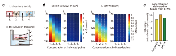
▲ Figure 2 c) Illustration of the structures of the existing culture platform and the microfluidic chip developed in the present study; d) The concentrations of the cytokines secreted from the microenvironmental cells over 24 hrs increased more rapidly and were maintained at higher levels in the microfluidic chip than in the existing culture platform; and. e) Of the substances secreted from the cells, Serpin E1, IL-8, and SPP-1 secretions were highest in the model developed by the present study.
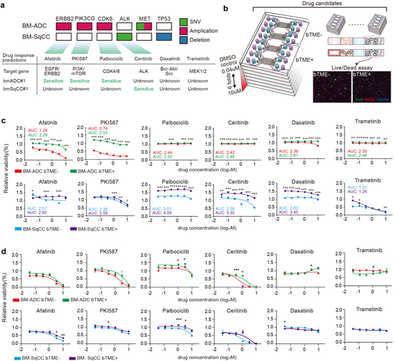
▲ Figure 3, a) The results of the genome-based prediction of the anticancer drug resistance of the patient-derived cancer cells; b) Schematic of drug evaluation using the developed microfluidic chip; c) The results of the drug evaluation performed in the developed microfluidic chip showed that the survival rate of the cancer cells co-cultured in the microenvironment increased; and d) The results of the drug evaluation performed in Transwell, the current culture platform, showed that no drug resistance caused by the environment was observed even under co-culture microenvironment conditions.